")

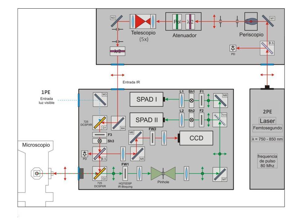
Nuestro grupo dispone de un microscopio de fluorescencia multi-fotónico y confocal láser de barrido que nos permite hacer estudios de micro-espectroscopía de fluorescencia con excitación de 1 fotón (1PE) o 2 fotones (2PE).
 |
 |
|
|
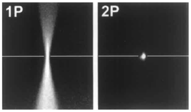
Excitación multifotónica (2PE) como alternativa a la microscopía confocal (1PE): alta resolución espacial sin necesidad de pinhole
| MICROSCOPÍA CONFOCAL (1PE): la fluorescencia emitida por la muestra se filtra mediante un pinhole colocado frente al detector, de manera que sólo se registra la fluorescencia emitida por el plano focal. |
|
MICROSCOPÍA MULTIFOTÓNICA (2PE): absorción simultánea de dos fotones de la mitad de energía (típicamente rojo o infrarrojo). Para ello se necesita localizar una gran cantidad de fotones de excitación en un punto determinado de la muestra (volumen confocal ~ 0.1 mm3) un objetivo de alta apertura numérica durante un tiempo muy corto, utilizando láseres pulsados de ~100 fs en el infrarrojo IR - IR cercano. |
 |
|
DIFERENCIAS 1PE vs 2PE: en 2P sólo se excitan los fluoróforos localizados en el foco: se minimiza el daño por la luz en las muestras y permite estudiarlas a profundidades mayores, mejorando la resolución de un microscopio confocal. FOTOTOXICIDAD GLOBAL REDUCIDA DE LA MUESTRA vs mayor FOTOBLANQUEO EN EL VOLUMEN CONFOCAL El micro-espectrofotómetro puede utilizar excitación 1PE o 2PE: la elección del tipo de excitación depende del tipo de muestra y de la fotofísica y concentración de fluoróforos. 2PE es menos sensible a la dispersión que 1PE, produciendo imágenes de mayor resolución y contraste. Además, permite tiempos de observación más largos para los estudios con célula viva, pero produce mayor fotoblanqueo y fototoxicidad en muestras delgadas con un bajo número de fluoróforos. En estos casos, 1PE funciona bien, ofreciendo una mejor resolución espacial. |
Time-resolved fluorescence spectroscopy through a microscope
In the last two decades, the techniques of confocal fluorescence microscopy have evolved to become essential tools to study complex cellular process. These technical advances have opened the door to fluorescence micro-spectroscopy studies, i.e. the possibility of quantitative photophysical and spectroscopic studies through a microscope with high spatial resolution (sub-μm), which can be extended to even single molecules.
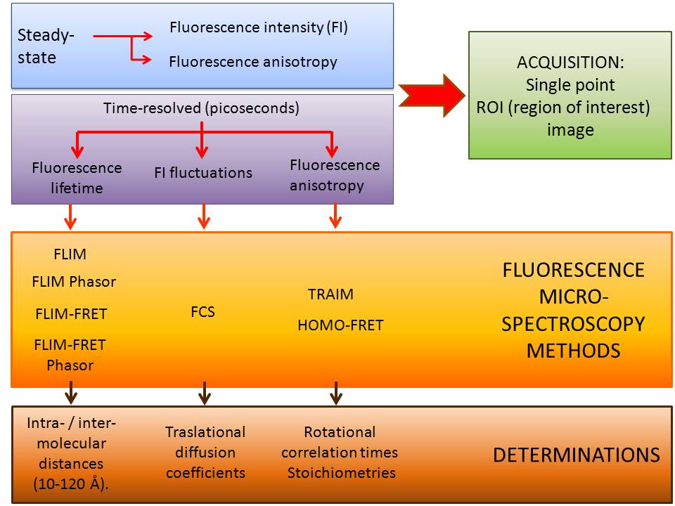
STEADY STATE AND TEMPORAL RESOLUTION MEASUREMENTS IN THE FLUORESCENCE MICRO-SPECTROMETER:
Micro-spectroscopy techniques are optimal to study complex physical-chemical processes in live cells and tissues, so they are minimally invasive: instead of fixing and physically sectioning a sample, it is possible to obtain a 3D dataset from a live specimen, through optical sectioning.
SAMPLES: IN VITRO OR IN VIVO (CELLULAR EXTRACTS, FIXED OR LIVING CELLS, TISSUES)
| NAME | STAFF | PHONE | |
| Mª Pilar Lillo Villalobos | Staff Scientist | 961027 | This email address is being protected from spambots. You need JavaScript enabled to view it. |
| Carolina García Rodríguez | Technical staff | 961201 | This email address is being protected from spambots. You need JavaScript enabled to view it. |
| Ulises Acuña Férnandez | Research Professor Ad honorem | 961220 | This email address is being protected from spambots. You need JavaScript enabled to view it. |
 |
 |
| FRET | ANISOTROPÍA DE FLUORESCENCIA |
 |
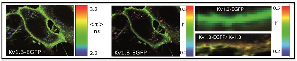 |
| IDENTIFICACIÓN DE ESPECIES MOLECULARES | ORGANIZACIÓN 3D |
 |
 |
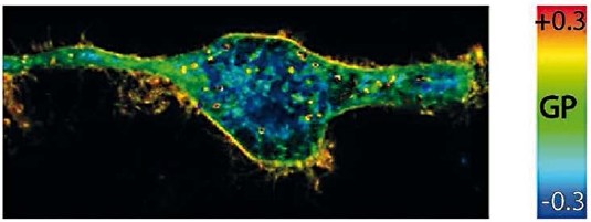
| MEMBRANA | FOTOFÍSICA |
 |
The overall objective of the Group is to understand how biological systems work under physiological conditions. With this aim we develop and implement both theoretical and experimental methods based on ps-resolved fluorescence spectroscopy and two-photon laser excitation microscopy, to provide the required temporal (ps-s) and spatial (submm- nm) resolution: fluorescence intensity (normalized desviation), fluorescence correlation spectroscopy (FCS), lifetime (FLIM, FLIM-phasors), energy transfer (FLIM-FRET, FLIM-phasors) and polarization fluorescence (TRAIM and homo-FRET). These are non-invasive quantitative methods that allow us to discriminate and characterize supramolecular structures in different subcellular locations, in very heterogeneous media, with single molecule resolution.
The applications include biomolecular conformational dynamics, organization and interactions of multiprotein complexes, and lipid microdomains distribution, in vitro, in living cells, and tissues.
STRATEGIC AIMS
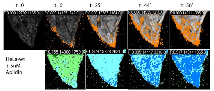
- Mechanism of action of bioactive compounds: Quantitative characterization of drug-target interactions in the cell membrane and inside the cell.
- Detection, identification, localization and quantification of in vivo cell markers.
- Detection of lipid micro-domains in model membrane systems and living cells, induced by bioactive compounds.
- 3-D organization, conformational dynamics and stoichiometry of multicomponent complexes.
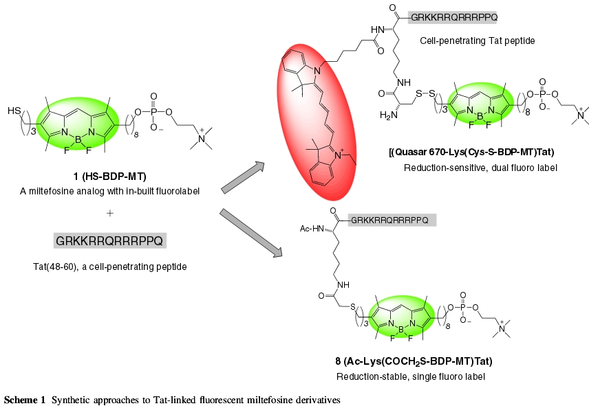
- Spectroscopic characterization of fluorescent materials for biotechnological applications.
- Fundamental Photophysics. Design, synthesis, characterization and applications of emitting molecular probes and fluorescent drugs.