Time-resolved fluorescence spectroscopy through a microscope
In the last two decades, the techniques of confocal fluorescence microscopy have evolved to become essential tools to study complex cellular process. These technical advances have opened the door to fluorescence micro-spectroscopy studies, i.e. the possibility of quantitative photophysical and spectroscopic studies through a microscope with high spatial resolution (sub-μm), which can be extended to even single molecules.
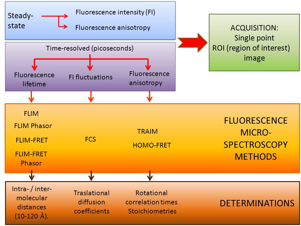
STEADY STATE AND TEMPORAL RESOLUTION MEASUREMENTS IN THE FLUORESCENCE MICRO-SPECTROMETER:
Micro-spectroscopy techniques are optimal to study complex physical-chemical processes in live cells and tissues, so they are minimally invasive: instead of fixing and physically sectioning a sample, it is possible to obtain a 3D dataset from a live specimen, through optical sectioning.
SAMPLES: IN VITRO OR IN VIVO (CELLULAR EXTRACTS, FIXED OR LIVING CELLS, TISSUES)